animal cell under microscope labeled
You should observe the cell membrane nucleus and cytoplasm Observation. The shape of animal cells also varies with some being flat others oval or rod-shaped.

Eukaryotic Cell Structure Animal Cell Plant And Animal Cells Eukaryotic Cell
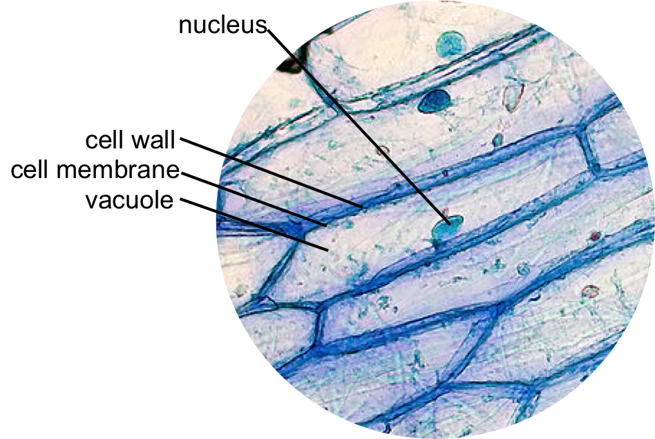
Onion Epidermis Under Light Microscope Purple Colored Large Cells Project Microscopic Photography Epidermis.

. Sunday April 18th 2021. Get more skin-labeled diagrams on social media for anatomy learners. We use microscope comprehensively in microbiology mineralogy cell biology biotechnology nano physics microelectronics pharmacology and forensics.
Hair under microscope. The following labeled drawings must be completed. When labeled with fluorescent dyes they are invaluable for locating specific molecules in cells by fluorescence microscopy Figure 9-15.
The granulated area is the cell Cytoplasm while the huge round part is the Nucleus. Within the epidermis of a skin you will find squamous diamond-shaped and polyhedral cells under the light microscope. Animal cells are smaller than the plant cells and they are generally irregular in shape taking various forms of shapes due to lack of the cell wall.
So hair is an epidermal down growth embedded into the dermis or hypodermis of the animals skin. Animal Cell Diagram Under Microscope. We all keep in mind that the human body is quite intricate and a method I.
They are all typical elements of a cell. Within the cell there is a shape of round with a circular structure of granulated part on the epithelial cells. We all keep in mind that the human physique is amazingly elaborate and one way I discovered to comprehend it is by way of the style of human anatomy diagrams.
When observing onion cells there is the Cell Surface Membrane which is present in all living cells. Most cells both animal and plant range in size between 1 and 100 micrometers and are thus visible only with the aid of a microscope. While observing with tissues or on tissue.
The neurons in the ventral horn of the spinal cord and cerebral cortex are the best examples of multipolar motor neurons. Illustrate only a plant cell as seen under electron microscope. Sep 15 2014 - Learn the structure of animal cell and.
The multipolar motor neuron under a microscope shows the typical features. Tuesday April 20th 2021. Observe the cheek cells under both low and high power of your microscope.
As stated before animal cells are eukaryotic cells with a membrane-bound nucleus. The animal cell is more fluid or elastic or malleable in structure. This neuron possesses the cell body an axon and several short branched dendrites.
Observing a wide range of biological processes and animal cell under light microscope is easier due to advances in microscopic techniques. These are both specific types of cells and from specific species. Multipolar motor neuron under microscope.
Here is an electron micrograph of an animal cell with the labels superimposed. Cell is a tiny structure and functional unit of a living organism containing various parts known as organelles. Thats the major difference between plant and animal cells under microscope.
The animal cell diagram is widely asked in Class 10 and 12 examinations and is beneficial to understand the structure and functions of an animal. The diagram is very clear and labeled. Animal Cell Diagram Under Microscope Labeled.
Animal cells are eukaryotic cells that contain a membrane-bound nucleus. A brief explanation of the different parts of an animal cell along with a. You see that many features are in common.
Hair under a compound microscope. The cylindrical shaft of the hair under a microscope shows three layers medulla cortex and cuticle of keratinized cells. Under the microscope animal cells appear different based on the type of the cell.
560 x 364 pixel electron microscope image animal cell and organelles labeled animal cell plasma membrane organelles. For viewing under the light microscope can label plant and animal cell structures and describe their functions to be able to work out the size of a cell contain chlorophyll which absorb light energy to make food by photosynthesis lipid membrane which controls what enters and leaves the cell space. Animal Cell Diagram Under Light Microscope.
Human cheek cells are made of simple squamous epithelial cells which are flat cells with a round visible nucleus that cover the inside lining of the cheekC. A typical animal cell is 1020 μm in diameter which is about one-fifth the size of the smallest particle visible to the naked eye. As you can see in the above labeled plant cell diagram under light microscope there are 13 parts namely Cell membrane.
Differences between animal and human hair. Labeled with electron-dense particles such as colloidal gold spheres they are used for similar purposes in the electron microscope discussed below. You can observe this epithelial animal cell under microscope with high power.
Labeled animal cell below electron microscope midbodyl. A cell is a very tiny structure which exists in living bodies. Illustrated in Figure 2 are a pair of fibroblast deer skin cells that have been labeled with fluorescent probes and photographed in the microscope to reveal their internal structure.
The largest animal cell is the ostrich egg which has a 5-inch diameter weighing about 12-14 kg and the smallest animal cells are neurons of about 100 microns in diameter. There are also more intriguing shapes such as curved spherical concave and rectangular. Heres a diagram of a plant cell.
Most of the cells are microscopic in size and can only be seen under the microscope. Draw a diagram of one cheek cell and label the parts. However the internal structure and organelles are more or less similar.
Observing plant cell or animal cell under microscope is important as a cell is a very small unit that cant be seen with your naked eye. This shows a generalized animal cell under a light microscope. Add a drop of purple stain specific for animals and cover with a cover slip.
It also shows the myoepithelial cells that surround each sweat gland of the animal skin. Skin cells under a microscope. They are different from plant cells in that they do contain cell walls and chloroplast.
Observe the cheek cells under both low and high power of your microscope. Hair can be matched by other characteristics that can be viewed under a compound microscope. The plant cell as more rigid and stiff walls.
Animal cell under the microscope. Function cell does in the body dictate the change and adaptation done by cell. The nuclei are stained with a red probe while the Golgi apparatus and microfilament actin network are stained green and blue respectively.
When you look at an animal or plant cell under a microscope the most obvious feature you will see is the large dark nucleus. The precise antigen specificity of antibodies makes them powerful tools for the cell biologist. Generalized Structure of a Plant Cell Diagram.
To make observations and draw scale. Our hair grows from follicles located under the skin and has two. You will find two main parts in hair a cylindrical shaft and a terminal hair follicle.

Cells And Dna Lesson Plan Science Cells Teaching Cells Middle School Science Activities

Animal Cell Model Diagram Project Parts Structure Labeled Coloring Plant And Animal Cells Animal Cell Animal Cells Model

Animal Cell Human Cell Diagram Animal Cell Drawing Cell Diagram

Muppets Animal Drawing At Paintingvalley Com Explore Collection Of Muppets Animal Drawing Cell Diagram Animal Cell Structure Animal Cell

Animal Cell Diagram Woo Jr Kids Activities Children S Publishing Cell Diagram Animal Cell Animal Cell Project

Epidermal Onion Cells Under A Microscope Plant Cells Appear Polygonal From The Cell Diagram Plant Cell Diagram Plant Cell

Labeled Animal Cell Diagram Cell Diagram Plant And Animal Cells Animal Cell Parts

Draw It Neat How To Draw Animal Cell Animal Cell Drawing Animal Cell Biology Diagrams

560 X 364 Pixel Electron Microscope Image Animal Cell And Organelles Labeled Animal Cell Plasma Membrane Organelles

Animal Cell Structure And Organelles With Their Functions Jotscroll Animal Cell Organelles Cell Diagram

Organelle Micrographs Google Search Teaching Biology Cell Biology Science Biology

Animal Cell Organelles Cell Organelles Organelles

Printable Labeled And Unlabeled Animal Cell Diagrams With List Of Parts And Definitions Animal Cells Model Animal Cell Cell Diagram

Histolab4a Htm Histology Slides Science And Nature Animal Cell

Label The Animal Cell Worksheets Sb11866 Animal Cells Worksheet Cells Worksheet Animal Cell

Animal Cell Organelles Sauna Design

Cell 8 Pictures Of Plant Cells Under A Microscope Plant Cell Structure Under Microscope Plant And Animal Cells Plant Cell Structure Plant Cell

Animal And Plant Cells Worksheet Inspirational 1000 Images About Plant Animal Cells On Pinterest Cells Worksheet Animal Cell Plant Cells Worksheet
